|
| ||||||
|
Urinary System - analyze the functional inter-relationships of the structures of the urinary system.
Chapter 38: Digestive and Excretory Systems Section 38.3 - The Excretory System Basic Renal Processes Renal Clearance |
Video: Section 38.3
|
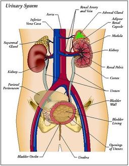
Urinary System (Click to Enlarge)
|
Urinary System
Your body takes nutrients from food and uses them to maintain all bodily functions including energy and self-repair. After your body has taken what it needs from the food, waste products are left behind in the blood and in the bowel. The urinary system works with the lungs, skin, and intestines—all of which also excrete wastes—to keep the chemicals and water in your body balanced.
Simply put, the urinary system is the organ system that produces, stores, and eliminates urine. In humans it includes two kidneys, two ureters, the bladder, and the urethra.
Did you know adults eliminate about a quart and a half of urine each day. The amount depends on many factors, especially the amounts of fluid and food a person consumes and how much fluid is lost through sweat and breathing.
Urinary System Anatomy
The kidney is the main organ of excretion, which is the process of removing nitrogen wastes from the body. Nitrogen compounds, mainly ammonia, are produced during the breakdown of proteins. Ammonia is very toxic, so it is quickly combined with carbon dioxide to produce urea. If left to accumulate, the body would be overwhelmed within days and the major organs would stop functioning. As a result, the kidney is one of the body’s essential organs. People whose kidneys fail must submit to artificial removal of wastes by dialysis, or have their kidneys replaced with a donated kidney.
As well as excretion, the kidneys regulate water concentration and pH (acid/base balance.) Additionally, the kidneys also release hormones important in Na+ regulation (renin) and red blood cell production (erythropoietin).
Labelling the Urinary System: online activity
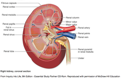
Kidney Anatomy and Urine Formation
Every day your body filters about 180 litres of water in the blood, which is the equivalent of a large oil drum. Obviously the fluids in the blood are filtered more than once, since the body’s total volume of blood is about 5 litres. From these 180 litres, about 2 litres of urine (approximately 1% of the volume being filtered) is produced each day. This varies as the kidneys balance water concentration, pH, and salt concentration.
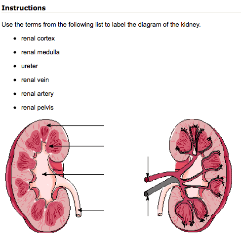
Study the following diagram of the anatomy of the kidney. You will be responsible for knowing these structures.
Every day your body filters about 180 litres of water in the blood, which is the equivalent of a large oil drum. Obviously the fluids in the blood are filtered more than once, since the body’s total volume of blood is about 5 litres. From these 180 litres, about 2 litres of urine (approximately 1% of the volume being filtered) is produced each day. This varies as the kidneys balance water concentration, pH, and salt concentration.
Study the following diagram of the anatomy of the kidney. You will be responsible for knowing these structures.
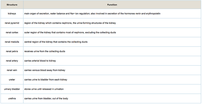
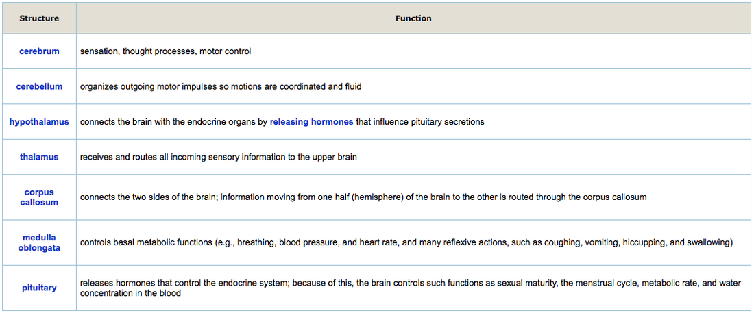
The functions of these structures are provided in the following table.
|
|
Nephron Structure
The nephron is the unit of filtration in the kidney. Each kidney contains at least a million nephrons. A nephron is composed of a capsule into which fluids and dissolved molecules and ions are squeezed under pressure. This fluid then passes through a series of tubules that selectively reabsorb molecules needed by the body, leaving the wastes in the fluid. Eventually the remaining fluid is expelled from the kidney in the form of urine. To fully understand this process, it is first necessary that you learn about the parts of the nephron.
In your Inquiry Into Life textbook, study Figure 16.4 on page 307. Take note of the location of the nephron in the kidney. Locate the glomerular capsule, also known as Bowman’s capsule. Trace the path of blood flow into the glomerulus. Notice how it enters through the afferent arteriole, passes through the glomerulus, and exits through the efferent arteriole. (Hint: it’s easy to remember the path of blood flow through these arterioles because they’re in alphabetical order.)
Study the names of the various parts of the nephron. You must be able to identify them from memory. (Vocabulary note: convoluted means twisted; proximal means near; distal means far; in this case, near and far refer to Bowman’s capsule)
Study Figure 16.4 on page 307 of the Inquiry Into Life textbook to see how the tubules are distributed within the renal medulla and cortex.
Redraw a nephron on a sheet of paper, label it, and keep it handy. You will need it later in the lesson. Label all the parts of the nephron on your diagram. Do not trace the diagram; draw it freehand. This will force your mind to remember the structures much better. Include the following: Bowman’s capsule, glomerulus, proximal convoluted tubule, loop of Henle, distal convoluted tubule, collecting duct.
Urine Formation
Urine formation has three stages—pressure filtration (also known as glomerular filtration), selective reabsorption, and tubular secretion.
Glomerular filtration
The Product of Nephron Function: UrineUrine is the ultimate product of nephron filtration and reabsorption. Urine is mostly water. The actual concentration of urine is determined by the relative concentration of water, which is varied under control according to the osmolarity (water balance) of the blood. You will have noticed the variation in colour of your own urine, which can be attributed to the amount of water you consume and the amount you lose due to evaporation. As well as water, urine contains:
On the drawing you made earlier in this lesson:
Urine formation has three stages—pressure filtration (also known as glomerular filtration), selective reabsorption, and tubular secretion.
Glomerular filtration
- Blood entering the kidney is under pressure. Blood enters the Bowman’s capsule and circulates through a loop of capillaries. Because it is under pressure, relatively small molecules, including water, salts, glucose, wastes, and amino acids, are filtered through the glomerular walls where the Bowman’s capsule absorbs them. Large molecules, such as proteins and blood cells, are left in the blood and circulate out to the peritubular capillary network.
- Glomerular filtrate passes out of Bowman’s capsule and passes through the tubules of the nephron, beginning with the proximal tubule near the Bowman’s capsule. Water from this dilute fluid moves by diffusion across the wall of the tubule and is reabsorbed by the peri-tubular capillary network. This diffusion is facilitated by the osmotic imbalance created by the highly concentrated blood emerging from the glomerulus.
This initial reabsorption increases the concentration of the urine in the tubule. Other small molecules in high concentration include glucose and amino acids. These are valuable resources, so they are actively reabsorbed, aided by carrier proteins in the cell in the membranes of the proximal tubule wall. The process is selective because the types of molecules reabsorbed are controlled by the specific carrier proteins.
- Tubular secretion occurs as molecules too large to be filtered into the glomerulus are actively transported across the wall of the distal convoluted tubule and into the urine. Molecules left over from muscle metabolism, hormone breakdown, and drugs such as antibiotics are removed from the blood in this way.
The Product of Nephron Function: UrineUrine is the ultimate product of nephron filtration and reabsorption. Urine is mostly water. The actual concentration of urine is determined by the relative concentration of water, which is varied under control according to the osmolarity (water balance) of the blood. You will have noticed the variation in colour of your own urine, which can be attributed to the amount of water you consume and the amount you lose due to evaporation. As well as water, urine contains:
- dissolved salts such as NaCl
- metabolic wastes, including nitrogenous wastes (i.e., urea and ammonia). These wastes are the result of protein breakdown that releases nitrogen compounds (i.e., ammonia). Ammonia is toxic, so is quickly converted to urea in the liver.
- small molecules resulting from the breakdown of hormones. This is the basis for urine testing of athletes. If irregular quantities of the products resulting from hormone breakdown are found in the urine, this may indicate the athlete has been taking supplements of these hormones, which are banned by most athletic regulatory bodies.
- Occasionally there will be unusual amounts of other products, such as protein or blood in the urine. These are not normal and usually signal something is not working properly. For example, high blood pressure may force proteins across the glomerular walls.
On the drawing you made earlier in this lesson:
- Indicate the direction of flow of fluids, beginning with glomerular filtration into Bowman’s capsule and ending with urine leaving the collecting duct.
- Indicate with arrows where each of the processes of urine formation occurs. Label the arrows.
| case_studies_in_renal_function.pdf |
Renal Artery vs. Renal Vein
You have learned about the functions of the kidney, the processes that occur there, and the hormones that are important in kidney function. Blood that flows into via the renal artery has a different composition than the blood flowing out of the kidney via the renal vein.
For this lesson you will complete a section assignment in which you will play the role of a new lab assistant in a lab where a mistake with patient records has been made. Your job is to resolve the problem accurately. The office manager, who risks being fired if the problem isn’t solved, has given you several documents and asked you to find a solution. Keep hard copies of these documents available while doing the assignment. Good luck!
There is no guided practice activity or glossary for this lesson.
Part 1
Write the final report on your findings. Indicate which patient (by number) is which. Give a brief explanation of why you made the identification. It doesn’t need to be long, just identify one characteristic of each pair of samples that led you to believe in their particular identification.
Remember, your job depends on getting it right. If you are correct, Les owes you one, big time!
Part 2
As a second part of the task, write a memo to Perry City explaining the request made to do the lab tests. When you first read Les’ memo, did it make you uneasy? Point out the ethical problems you have with the request made by Les, and identify as many breaches in ethical behaviour as you can.
You can find information about business ethics on the Biology 12 Web site Lesson 4.2C Renal Artery vs. Renal Vein.
Do you feel that Aldo handled the request appropriately? Explain your answer in a few sentences.
Click on the PDF link below this one to read the case studies.
|
| ||||
|
Nervous System - analyze the transmission of nerve impulses, analyze the functional inter-relationships of the divisions of the nervous system.
Chapter 35: Nervous System Section 35.1 - Human Body Systems Section: 35.2 - The Nervous System Section 35.3 - Divisions of the Nervous System Section 35.4 - The Senses Section 35.5 - Drugs and the Nervous System |
Videos: Section 35.2
|
Song Intro Youtube
Nervous System (Click to Enlarge)
|
|
Nervous System
Tasting, smelling, seeing, hearing, thinking, dreaming, breathing, heart beating, moving, running, sleeping, laughing, singing, remembering, feeling pain or pleasure, painting, writing...you couldn’t do any of these things without your nervous system! Your nervous system is made up of your brain, your spinal cord, and an enormous network of nerves that thread throughout your body, it’s the control center for your entire body. Your brain uses information it receives from your nerves to coordinate all of your actions and reactions. Without it, you couldn’t exist! |
Neurons—Electric Cells
As you read this lesson, your nervous system is performing a complex series of electrical events, all propagated along neurons. Neurons, along with neuroglial cells, are specialized cells that make up the nervous system.
The nervous system is divided into two functional divisions—the peripheral nervous system (PNS), made up of motor and sensory neurons, and the central nervous system (CNS). Neurons are found in both systems. These two systems will be explored in future lessons.
There are three kinds of neurons: motor neurons, sensory neurons, and interneurons.
- Motor neurons carry impulses (individual electrical events called action potentials) from the central nervous system (brain and spinal cord) to muscles and organs (i.e., the heart)
- Sensory neurons carry impulses from the sensory receptors (any cell that detects external or internal stimulation, such as those in the retina of the eye that detect light) to the central nervous system
- Interneurons carry impulses from sensory neurons to motor neurons, and are located solely in the central nervous system
|
|

|
Neuron Structure
Throughout the body, neurons are organized in different ways, and their structures and functions are subtly different. The following questions will help you focus on their differences and similarities. Use the diagram on page 321 in your Inquiry Into Life textbook for comparison.
In the drawing, locate the cell bodies. Now note the extensions from the cell bodies. At the top of the illustrations, you’ll see that the extensions are either axon or dendrites. The extensions at the bottoms of each illustration are all dendrites.
The arrows indicate the direction an impulse travels along the neuron. In all cases, it’s from the top to the bottom of the cell.
The movement of the impulse in relation to the extensions can be used to write a general rule about the direction of impulse travel in neurons:
Impulses travel from dendrites toward the cell body and then down the axons.
Use the diagrams in your Inquiry Into Life text and use this rule to trace the path of an impulse from receptor to effector. Effectors are located at the end of motor neurons. They respond to impulses that travel down the axons of motor neurons.
Label the Neuron Structure: online activity
Label the Neuron: online activity 2
Myelin Sheath
The myelin sheath is an important component of neuron structure. Cells that have a myelin sheath are described as myelinated. Myelinated axons enable the neuron to quickly transfer or propagate an impulse. In the human body, myelinated axons carry impulses at about 2 metres/second. Non-myelinated dendrites carry impulses about 50 to 100 times more slowly than myelinated axons.
Myelinated axons are found in long nerves that connect sensory organs, for example, the skin on your toes, to the central nervous system. A disease called multiple sclerosis (MS) attacks the protective myelin covering of neurons, and sometimes destroys it. This damage interrupts the normal movement of nerve impulses along the axons. The symptoms of MS vary greatly from person to person, ranging from problems with vision and coordination to partial or even complete paralysis.
| neuron_comparison_ws.doc |
Nerve Impulse Transmission
For the human body to work properly, it must be able to gather external and internal information, send the information to a processing centre to be deciphered, and send appropriate responses to various muscles and organs. This is largely accomplished by neurons “firing” in a controlled and predictable manner. The biochemical events that occur inside and along the surface of neurons, called the action potential, is the primary means of sending information through the body. In this lesson you will learn how the neuron manages this complex and yet simple process, and how some neurons increase the speed of impulse travel.
The Resting Membrane Potential
Individual neurons carry impulses in only one direction, and from a particular source towards the central nervous system. All neurons work exactly the same way. When you accidentally bump your eye, you see “stars” or tiny pinpricks of light. The bump caused neurons that normally carry light messages to send a message to the brain, and the brain only interprets messages from this part of the body as light.
To understand how a neuron carries an impulse, we need to understand how various ions move around in the neuron.
Scientists have learned a great deal about neurons by studying squid, because these animals have extremely large axons—in some cases, the size of a pencil. Small electrical probes (microprobes) can be placed inside and along the surface of these large neurons.
Scientists had hypothesized that electrical events along the neuron’s surface were responsible for carrying impulses. By carrying out these experiments with squid, they were able to observe electrical potentials and changes while neurons were both at rest and carrying an impulse.
At rest, axon surfaces have a tiny electrical potential of –65 millivolts (a single dry cell has an electrical potential, from one end to the other, of 1500 millivolts or 1.5 volts) between the outside and the inside of the membrane
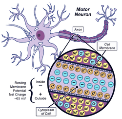
The diagram shows a motor neuron with a shortened axon. Notice that one piece of the axon is redrawn in the bottom of the diagram. Pay particular attention to:
Questions to Consider:
- the location of the inside and outside of the membrane
- the distribution of K+, Na+, and organic anions
- the relative number of each ion (note the uneven balance between positive and negative ions outside, and then inside)
- the potential difference (measured in millivolts) between the outside and the inside of the membrane, shown on the right of the drawing
Questions to Consider:
- Which ions are important in the resting membrane potential?
- What is the potential difference between the outside and inside of a neuron at resting membrane potential?
- Is conduction along an axon caused by a chemical or electrical phenomenon?
The Action Potential
When at rest, a neuron’s membrane maintains an uneven distribution of ions. When a receptor is stimulated (e.g., light in the retina, pressure on fingertip, etc.) above the threshold level, the neuron goes into action potential. To understand the events taking place in the neuron, more data were obtained by studying squid membranes as they actually carried impulses. As impulses travelled past microprobes, the electrical potential varied. When this data is presented in a graph, it looks like this:
When at rest, a neuron’s membrane maintains an uneven distribution of ions. When a receptor is stimulated (e.g., light in the retina, pressure on fingertip, etc.) above the threshold level, the neuron goes into action potential. To understand the events taking place in the neuron, more data were obtained by studying squid membranes as they actually carried impulses. As impulses travelled past microprobes, the electrical potential varied. When this data is presented in a graph, it looks like this:
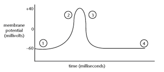
Here’s what happened.
- The membrane is at resting membrane potential.
- As an impulse passes the microprobe, the potential reverses to +40 millivolts (this is called depolarization).
- As the impulse moves on, the membrane returns to resting potential (this is called repolarization).
- During the period after repolarization, the membrane is in a refractory period.
Events Inside the Axon
How can events inside the axon change the ion distribution to account for these changes in potential?
How can events inside the axon change the ion distribution to account for these changes in potential?
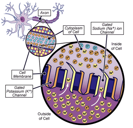
Notice in this diagram that the membrane has two types of protein molecules called gated channels. One allows the controlled movement of Na+, and the other K+. Since the distribution of Na+ at RMP is greater on the outside, opening the Na+ gates allows the Na+ to rush in by diffusion. This occurs at the beginning of the stage called action potential (AP), the series of events that occur as an impulse travels past a point on the membrane. The inward rush of Na+ causes a higher distribution of positive charges on the inside, which accounts for the reverse in polarity (to +40mV) across the membrane. (depolarization)
Immediately after the Na+ gates open, the K+ gates open and potassium rushes out, again by diffusion. This causes the polarity of the membrane to revert to its RMP (repolarization).
To regain true RMP, ion distribution the Na+ and K+ must be reversed. To do this, the membrane uses another protein molecule—a Na+/K+ pump. These molecules pick up the misplaced ions and, with the expenditure of ATP, the molecule changes shape and pumps the ions back into their normal RMP configuration. The time it takes to recover RMP is known as the refractory period. During RMP the Na+/K+ pump works to maintain the imbalance of ions characteristic of resting membranes.
Note:This use of ATP is an active process, which means energy must be used up in the process. This prevents impulses from travelling in both directions—an important feature of axons.
Go to the Biology 12 Web site Lesson 4.1B Impulse Transmission:
Action Potential Details
The time taken for an action potential to pass one site on a membrane is about 4 milliseconds. At maximum capacity, this amounts to 250 impulses per second. The actual number is a bit lower because there are inefficiencies in delivering ATP to any one site and that slows the process.
Since the neuron’s capacity to carry impulses is limited, increased stimulation of a neuron beyond this is not noticed. This means that once you have applied a maximum amount of pressure to your fingertip, pressing harder will not cause the brain to produce a sensation of more pressure.
Greater stimulation, or increased pressure, is translated as more axons firing, rather than one axon firing harder. Also, below a certain stimulus a neuron will not fire. This is known as a threshold. This important principle in action potential is called the all-or-none response. The neuron either reaches threshold stimulation and fires (initiates an impulse), or it doesn’t reach threshold stimulation and nothing happens. Individual neurons cannot vary the way they carry action potentials. For variation in sensation to occur, various numbers of neurons fire. Hitting your thumb with a hammer causes a huge number of neurons to fire. Gently touching the hammer only stimulates a few. The brain interprets the number of neurons firing as variation in stimulation.
Propagation of Action Potential
Let’s consider what happens at a particular site on a neuron when it is in AP. The disruption of ion balance at that site stimulates the adjacent membrane (in RMP). Specifically, as Na+ ions leak into the cell’s cytoplasm in AP, Na+ ions from the adjacent part of the membrane diffuse in to replace them. This weakens the RMP at that site, which in turn stimulates the membrane at that site to go into AP. The wave of AP down the neuron is the moving impulse. As you can imagine, this process is too slow to account for the high speed of impulse travel within the nervous system. Consider how long it would take for information from the tail of a whale to reach its brain. Far too long to allow for its graceful movements.
Action Potential Animation
Immediately after the Na+ gates open, the K+ gates open and potassium rushes out, again by diffusion. This causes the polarity of the membrane to revert to its RMP (repolarization).
To regain true RMP, ion distribution the Na+ and K+ must be reversed. To do this, the membrane uses another protein molecule—a Na+/K+ pump. These molecules pick up the misplaced ions and, with the expenditure of ATP, the molecule changes shape and pumps the ions back into their normal RMP configuration. The time it takes to recover RMP is known as the refractory period. During RMP the Na+/K+ pump works to maintain the imbalance of ions characteristic of resting membranes.
Note:This use of ATP is an active process, which means energy must be used up in the process. This prevents impulses from travelling in both directions—an important feature of axons.
Go to the Biology 12 Web site Lesson 4.1B Impulse Transmission:
- to see a short animation of this process.
- to see a detailed animation of the action potential.
Action Potential Details
The time taken for an action potential to pass one site on a membrane is about 4 milliseconds. At maximum capacity, this amounts to 250 impulses per second. The actual number is a bit lower because there are inefficiencies in delivering ATP to any one site and that slows the process.
Since the neuron’s capacity to carry impulses is limited, increased stimulation of a neuron beyond this is not noticed. This means that once you have applied a maximum amount of pressure to your fingertip, pressing harder will not cause the brain to produce a sensation of more pressure.
Greater stimulation, or increased pressure, is translated as more axons firing, rather than one axon firing harder. Also, below a certain stimulus a neuron will not fire. This is known as a threshold. This important principle in action potential is called the all-or-none response. The neuron either reaches threshold stimulation and fires (initiates an impulse), or it doesn’t reach threshold stimulation and nothing happens. Individual neurons cannot vary the way they carry action potentials. For variation in sensation to occur, various numbers of neurons fire. Hitting your thumb with a hammer causes a huge number of neurons to fire. Gently touching the hammer only stimulates a few. The brain interprets the number of neurons firing as variation in stimulation.
Propagation of Action Potential
Let’s consider what happens at a particular site on a neuron when it is in AP. The disruption of ion balance at that site stimulates the adjacent membrane (in RMP). Specifically, as Na+ ions leak into the cell’s cytoplasm in AP, Na+ ions from the adjacent part of the membrane diffuse in to replace them. This weakens the RMP at that site, which in turn stimulates the membrane at that site to go into AP. The wave of AP down the neuron is the moving impulse. As you can imagine, this process is too slow to account for the high speed of impulse travel within the nervous system. Consider how long it would take for information from the tail of a whale to reach its brain. Far too long to allow for its graceful movements.
Action Potential Animation
Saltatory Conduction
To allow for quick impulse travel (up to 700 kilometres per hour) impulses actually jump along the neuron. Refresh your memory of axon structure with this diagram.
To allow for quick impulse travel (up to 700 kilometres per hour) impulses actually jump along the neuron. Refresh your memory of axon structure with this diagram.

Notice the structures labelled X in the diagram. These are Schwann cells, which collectively produce a sheath called the myelin sheath. The little gaps that interrupt the Schwann cells are called the nodes of Ranvier.
|
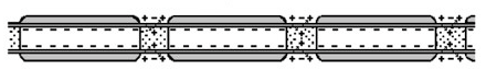
The Na+ and K+ gates in myelinated axons are concentrated at these nodes. As an impulse travels along the neuron, it actually jumps from node to node, by a process called saltatory conduction. This jumping accounts for the great speed of conduction in long axons.
Click on How Does an Action Potential Occur? to view an animation illustrating how an action potential is set up in a neuron |
Synaptic Transmission
Impulse transmission occurs in two ways—neuron transmission, which is electrochemical, and synaptic transmission, which is chemically controlled. Synaptic transmission takes place at the gaps between neurons, or between neurons and effectors. Synaptic transmission is controllable, which means the impulse can be tuned or ignored. Synaptic transmission is influenced by neurohormones, such as adrenalin or drugs that can mimic or enhance the effect of natural neurotransmitters. Common drugs such as aspirin, and opiates, including cocaine or morphine, work in this way.
Structure of Synapses
Synapses are small gaps that separate communicating neurons or motor neurons from effector organs. Impulses travelling along neurons must first cross these gaps before they cause the next (postsynaptic) cell to be stimulated.
Open your Inquiry Into Life textbook to Figure 17.5 on page 324.
In the diagram in the top left corner, notice the axon of a sensory neuron and the entire interneuron. Normally the interneuron is connected to other interneurons, or perhaps to motor neuron cell bodies.
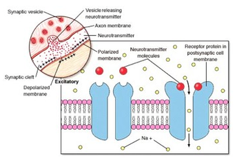
In the diagram in the top right corner, you’ll notice the swelling at the end of the axon. This is called the axon bulb. The axon bulb contains vesicles loaded with protein molecules called neurotransmitters. The two neurons do not touch each other. Instead they are separated by a tiny gap called the synaptic cleft. If the postsynaptic cell is a muscle cell, then the gap is called a neuromuscular junction.
You should also check out the diagrams in the Essential Study Partner on the Biology 12 Web site Lesson 4.1 C Synaptic Transmission. Compare these diagrams to those of the synapse structure on page 324 in the Inquiry Into Life textbook.
The following electron micrograph shows several neurons in the brain and the synapses between them. The synapses are labelled. Notice the scale at the top right of the photo (0.5 µm is 5 x 10⁻⁷ m). The arrowheads indicate the membrane of one neuron. The “thorns” are the axon bulbs. Notice the synaptic vesicles that contain neurotransmitter molecules inside.
How Synapses Work
An action potential travels along a neuron. When it reaches the axon bulb, it causes the bulb membrane to increase its calcium ion (Ca++) permeability. This causes Ca++ to rush into the axon bulb, where vesicles containing neurotransmitters are located. These vesicles then fuse with the membrane, causing the neurotransmitter molecules to be released into the cleft. Once released, they quickly diffuse across the tiny cleft to the postsynaptic cell membrane where receptors embedded in the postsynaptic membrane attach to the neurotransmitter. These receptors are actually Na+ gates, which are opened by the presence of the neurotransmitter. Once open, Na+ rushes into the cell, causing the postsynaptic cell to go into action potential. The postsynaptic cell then carries the impulse along its length.
Impulse transmission occurs in two ways—neuron transmission, which is electrochemical, and synaptic transmission, which is chemically controlled. Synaptic transmission takes place at the gaps between neurons, or between neurons and effectors. Synaptic transmission is controllable, which means the impulse can be tuned or ignored. Synaptic transmission is influenced by neurohormones, such as adrenalin or drugs that can mimic or enhance the effect of natural neurotransmitters. Common drugs such as aspirin, and opiates, including cocaine or morphine, work in this way.
Structure of Synapses
Synapses are small gaps that separate communicating neurons or motor neurons from effector organs. Impulses travelling along neurons must first cross these gaps before they cause the next (postsynaptic) cell to be stimulated.
Open your Inquiry Into Life textbook to Figure 17.5 on page 324.
In the diagram in the top left corner, notice the axon of a sensory neuron and the entire interneuron. Normally the interneuron is connected to other interneurons, or perhaps to motor neuron cell bodies.
In the diagram in the top right corner, you’ll notice the swelling at the end of the axon. This is called the axon bulb. The axon bulb contains vesicles loaded with protein molecules called neurotransmitters. The two neurons do not touch each other. Instead they are separated by a tiny gap called the synaptic cleft. If the postsynaptic cell is a muscle cell, then the gap is called a neuromuscular junction.
You should also check out the diagrams in the Essential Study Partner on the Biology 12 Web site Lesson 4.1 C Synaptic Transmission. Compare these diagrams to those of the synapse structure on page 324 in the Inquiry Into Life textbook.
The following electron micrograph shows several neurons in the brain and the synapses between them. The synapses are labelled. Notice the scale at the top right of the photo (0.5 µm is 5 x 10⁻⁷ m). The arrowheads indicate the membrane of one neuron. The “thorns” are the axon bulbs. Notice the synaptic vesicles that contain neurotransmitter molecules inside.
How Synapses Work
An action potential travels along a neuron. When it reaches the axon bulb, it causes the bulb membrane to increase its calcium ion (Ca++) permeability. This causes Ca++ to rush into the axon bulb, where vesicles containing neurotransmitters are located. These vesicles then fuse with the membrane, causing the neurotransmitter molecules to be released into the cleft. Once released, they quickly diffuse across the tiny cleft to the postsynaptic cell membrane where receptors embedded in the postsynaptic membrane attach to the neurotransmitter. These receptors are actually Na+ gates, which are opened by the presence of the neurotransmitter. Once open, Na+ rushes into the cell, causing the postsynaptic cell to go into action potential. The postsynaptic cell then carries the impulse along its length.
From Inquiry into Life, 9th Edition - Essential Study Partner CD-Rom. Reproduced with permission of McGraw-Hill Education
In the illustration, notice how the presence of the neurotransmitter alters the shape of the receptor molecules, allowing the Na+ to move into the cell. Remember that the shape of protein molecules is a factor of how they functioning. This movement initiates an action potential.
In the illustration, notice how the presence of the neurotransmitter alters the shape of the receptor molecules, allowing the Na+ to move into the cell. Remember that the shape of protein molecules is a factor of how they functioning. This movement initiates an action potential.
| synapse_diagram.pdf |
|
Instructions
Click on the pdf to label the diagram using the provided terms.
|
|
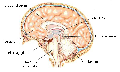
The Brain
The brain is the most complex organ in the body. It is responsible for our thoughts, memory, and emotions, and it controls functions such as hormone secretions by organs throughout the body. In this lesson you will become more familiar with some of the basic structures and functions of the brain.
Before we can discuss how the brain works, you need to familiarize yourself with its structure. Begin by identifying the structures in this chart on the labelled diagram that accompanies it. As you identify each structure on the diagram, read its function.
|
|
|
From Inquiry into Life, 9th Edition - Essential Study Partner CD-Rom. Reproduced with permission of McGraw-Hill Education
Read the section titled The Brain starting on page 328 in the Inquiry Into Life textbook. The glossaries at the end of this lesson and in the Inquiry Into Life textbook will also help you become familiar with the structures of the brain.
Now go to the Biology 12 Web site Lesson 4.1 F The Brain to review the brain’s structure. For another look at the structure of the brain, take a look at this virtual fetal pig dissection. Click on the link and review the entire dissection of the nervous system.
You can also use a search engine and the key words “brain structures” to find images of the brain on the Internet.
Neuro-Endocrine Control
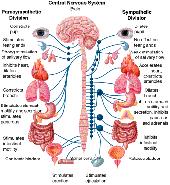
The brain is responsible for direct nervous control of muscles, memory, thought processes, emotions, and sensory awareness. It also controls the body by directing the release of hormones. The centre of this control is in the hypothalamus and the pituitary gland.
The hypothalamus receives information from the brain and monitors hormone levels in the blood. It responds to this information by releasing the appropriate hormones, which are then transported to the pituitary via axons that connect the two parts of the brain. Both are called endocrine glands because they produce hormones that are released directly into the blood stream and carried to a particular type of tissue. Some hormones control a broad range of tissues and others are very specific.
Take a look at the diagram of the pituitary gland in Figure 20.2 on page 397 of your Inquiry Into Life textbook. Notice that the pituitary had two portions—the anterior pituitary (closer to front of the brain) and the posterior pituitary. The function of anterior pituitary is more diverse than that of the posterior pituitary.
Now go to the Biology 12 Web site Lesson 4.1 F The Brain to review the brain’s structure. For another look at the structure of the brain, take a look at this virtual fetal pig dissection. Click on the link and review the entire dissection of the nervous system.
You can also use a search engine and the key words “brain structures” to find images of the brain on the Internet.
Neuro-Endocrine Control
The brain is responsible for direct nervous control of muscles, memory, thought processes, emotions, and sensory awareness. It also controls the body by directing the release of hormones. The centre of this control is in the hypothalamus and the pituitary gland.
The hypothalamus receives information from the brain and monitors hormone levels in the blood. It responds to this information by releasing the appropriate hormones, which are then transported to the pituitary via axons that connect the two parts of the brain. Both are called endocrine glands because they produce hormones that are released directly into the blood stream and carried to a particular type of tissue. Some hormones control a broad range of tissues and others are very specific.
Take a look at the diagram of the pituitary gland in Figure 20.2 on page 397 of your Inquiry Into Life textbook. Notice that the pituitary had two portions—the anterior pituitary (closer to front of the brain) and the posterior pituitary. The function of anterior pituitary is more diverse than that of the posterior pituitary.
Feedback Loop
The brain’s neuro-endocrine control of the body is carried out by the release of hormones. Short-term concentrations of hormones are adjusted by a mechanism called a feedback loop. Most homeostatic control operates in this way.
Recall that in feedback loops, an alteration of normal conditions will set off a string of connected events that eventually lead to the system being rebalanced. Feedback loops are usually negative because the body is working to reverse a condition that is out of balance.
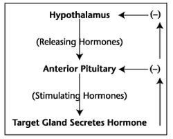
This flowchart summarizes a feedback loop in which the hypothalamus first produces a releasing hormone (RH), under direction of the brain. This RH causes the anterior pituitary to release a stimulating hormone (SH), which in turn causes a target gland to produce a particular hormone. This hormone has an inhibitory affect on both the hypothalamus and the anterior pituitary, slowing the output of each respective hormone. This is negative feedback because it inhibits production. In a few cases, a feedback loop can be positive. This will be discussed in a later lesson.
| nervous_system_written_response.pdf |
| fetal_pig_dissection.pdf |
|
Reproductive System - analyze the functional inter-relationships of the structures of the male reproductive system, analyze the functional inter-relationships of the structures of the female reproductive system.
Chapter 39: Endocrine and Reproductive Systems Section 39.1 - The Endocrine System Section 39.2 - Human Endocrine Glands Section 39.3 - The Reproductive System Section 39.4 - Fertilization and Development |
Video: Section 39.2
|
Reproduction
The major function of the reproductive system is to ensure survival of the species. Other systems in the body, such as the nervous and urinary systems, work continuously to maintain homeostasis for survival of the individual. An individual may live a long, healthy, and happy life without producing offspring, but if the species is to continue, at least some individuals must produce offspring.
This final section in the course focuses on both male and female reproductive systems and within each, what factors may affect them.
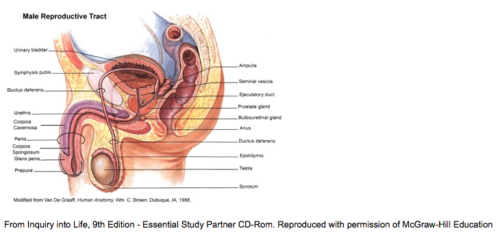
Male Reproductive Anatomy
Human reproduction is a complex process, as you are no doubt aware. The brain is tuned to behave and respond to others, often without our being aware or it. It determines, and to a large extent this affects how we interact socially. In this course you are only responsible for understanding the human reproductive system and the hormones that control it. The effect of pregnancy on hormone levels will be briefly discussed, but the course does not cover fetal development.
Male Reproductive AnatomyBefore you begin your study of human reproduction, you need to be familiar with the anatomy of the male and female reproductive systems. This lesson focuses on male reproductive anatomy. As you read the following descriptions, refer to the diagram in this lesson and the one in your Inquiry Into Life textbook on page 416.
External AnatomyThe primary function of the penis is to deposit sperm inside the vagina of a female during copulation. It also contains the final few centimetres of the urethra.
The scrotum is a sac consisting of skin, with embedded muscles, that contains the testes. The scrotum plays an important role in temperature regulation. As body temperature drops, the muscles in the wall of the scrotum contract, pulling the testes up against the body wall and helping them to retaining heat. Sperm formation in the testes will only take place at a few degrees below body temperature, so the testes are suspended outside the body in order for this to happen.
Internal AnatomyThe testes produce sperm and the male reproductive hormone testosterone. In the next lesson you will study sperm formation in more detail.
The epididymis is a folded tube in which sperm spend time acquiring their tails and maturing, which is required for them function properly.
The ductus deferens (vas deferens) is the tube that carries sperm from the epididymis through a circuitous route through the body wall, the prostate, and finally the urethra.
The prostate (not prostrate, which means to lay flat) is a gland that produces secretions that buffer the acidic environment of the vagina, assuring that a hostile environment does not destroy the sperm. Prostate cancer or enlargement afflicts many older men, making urination difficult. Regular prostate exams as men approach middle age can detect prostate cancer, increasing the possibility of early detection.
A pair of seminal vesicles flanks the prostate. Seminal vesicles produce fluids, including fructose, to fuel sperm as they move through the female reproductive system. The hormone prostaglandin is also produced there. Prostaglandins have a powerful but short-lived effect on muscles, causing them to contract. They act on the wall of the vagina and uterus, causing them to contract, which allows the passage of sperm into the uterus. This enhances the possibility of fertilization.
Cowper’s gland (bulbourethral) is a small gland that secretes lubricants to assist the sperm in their journey. Recent studies suggest these may produce antibodies against sperm from other individuals. Other species also have methods for preventing fertilization from competing males.
Go to the Biology 12 Web site Lesson 4.3A Male Reproductive Anatomy to read a scholarly article about this ability in birds.
The reproductive and urinary systems share a tube called the urethra. During copulation, it carries seminal fluid out of the body during orgasm. It also carries urine during urination.
The male gametes are called sperm.
The seminal fluid (semen) is made up of sperm and the secretions from various reproductive glands. If a male is sterile, sperm are produced in insufficient numbers to cause fertilization. If the vas deferentia are tied artificially (vasectomy), seminal fluid is produced, but without sperm.
|
|
This diagram is similar to the one on page 416 in your Inquiry Into Life text. It is a mid-saggital section. Some organs are off centre, and technically don’t belong in this view. The diagram in your Inquiry Into Life text also includes a view from the front, so you can see which organs are in this category.
Sperm Anatomy and the Pathway of Seminal FluidSperm may seem like simple structures whose only job is to carry DNA from the male to the female, but they are more complex than that. In this lesson you will learn to recognize and name the anatomy of sperm, and describe the path of sperm as it leaves the body.
Sperm Anatomy
Sperm cells are present in almost all plant and animal species, and their dependency on a watery environment has persisted through time. This almost always requires the sperm to have the ability to swim, so the cells have a tail. In some species, sperm cells have more than one tail. As well there is a “motor”’ that contains mitochondria, which provide ATP used in the flagellum to change shape, or beat in a characteristic motion reminiscent of fish or amphibians.
Keep in mind that a sperm cell is a single cell, and its cellular complexity approaches that of some protists. They are short lived. Once released from the body, their life expectancy is only about two days. This means they must be placed inside the female within two days of ovulation for fertilization to occur.
Study Figure 21.3 on page 418 of your Inquiry Into Life textbook.
The Pathway of Sperm
The pathway of sperm as it leaves the body is straightforward. From the testes, where sperm are produced, the cells travel to the epididymis where they mature. From there, during orgasm, muscular contractions of the epididymis and the vas deferens propel the sperm out of the vas. As they move, they pass several organs, each of which contributes to the seminal fluid. Without these fluids, sperm are incapable of fertilizing an egg. Sperm first pass the seminal vesicles, then the prostate, and finally the Cowper’s glands. The final pathway of sperm is the urethra, which leads out of the body.
Several malformations, either natural or artificial, can prevent sperm from moving through this pathway. Most notable are vasectomy and scarring, which is caused by bacterial infections of the inner lining of the tube. Sexually transmitted diseases, such as syphilis and gonorrhea, produce scarring and reduce fertility.
Note that reduced fertility means there is a reduced chance of producing a fertilized egg (pregnancy). Sterility means the chance is zero.
Go to the Biology 12 Web site Lesson 4.3B Sperm Anatomy and the Pathway of Seminal Fluid for more information about reduction in sperm cell counts. Also review “Introduction, Human Male System and Male Anatomy.”
Behaviour of Sperm
Depending on a man’s physical condition, sperm may be weak swimmers or malformed, often having multiple tails. Some sperm appear to be more interested in fighting than uniting. When sperm from multiple men is mixed together in a vagina, sperm cells compete with one another. This isn’t surprising, since it has only been relatively recently, geologically speaking, that mixing sperm was not the norm. Consider the evolutionary advantage of a male producing the best fighting sperm. While engaging the opponent’s sperm with a battle royal, others from the same man could be successfully fertilizing the egg. Once fertilized the egg accepts no other sperm.
The Testes Produce Sperm and Hormones
The testes have two roles in reproduction: to produce sperm cells and to produce hormones.
Sperm cell production, called spermatogenesis, occurs in the seminiferous tubules. Sperm are gametes that result from the cell division process called meiosis. Meiosis occurs continuously, from puberty to death, and the process requires a temperature just slightly lower than body temperature.
Hormones are also produced in the testes. Testosterone production occurs in the interstitial cells and inhibin is produced in the seminiferous tubules. Hormone secretions are regulated by the anterior pituitary and the hypothalamus, and controlled by negative feedback loop. The various sex hormones have several functions.
Testosterone
- controls development of primary sex organs
- controls development of secondary sex characteristics, beginning at onset of puberty: growth of body hair, enlargement of vocal cords (causes voice to deepen), growth of adult physique; later it controls receding hair line in male pattern baldness
- released by hypothalamus
- stimulates the anterior pituitary to release two controlling hormones (LH and FSH)
- stimulates production of sperm
- also known as interstitial cell stimulating hormone—ICSH
- stimulates production of testosterone
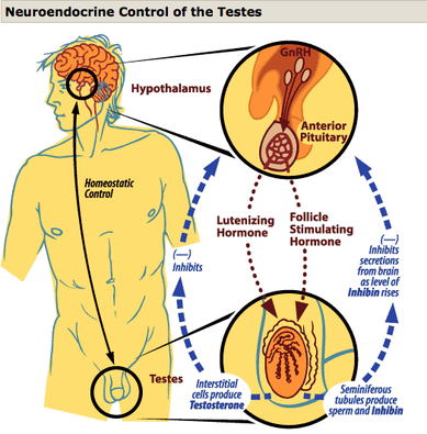
Study the diagram that shows neuroendocrine control of the testes and note the following characteristics:
- This is a negative feedback loop. Some event, e.g., secretion of LH, causes something to happen (testosterone secretion), which in turn reduces the amount of LH released. This allows hormone levels to be self-regulating.
- Before puberty, very low levels of FSH and LH are released from the brain, and this suppresses sexual development.
- Ultimately the brain is in control of sexual functioning.
- Hormones circulate in the bloodstream, so levels can be monitored. It is possible to introduce artificial levels of hormones that would bring about designed changes in the body, but some side effects would be unpleasant. Read the last paragraph in Hormonal Regulation in Males on page 419 in your Inquiry Into Life textbook.
| reproductive_system_written_response.pdf |
Female Reproductive Anatomy
This lesson focuses on the basic reproductive anatomy and processes of human females. You may be surprised to learn that in spite of the obvious physical differences between males and females, the development of sexual structures is quite similar.
Anatomy of the Female Reproductive System
At the most basic level the reproductive system performs the following functions:
Internal Anatomy
This is a mid-saggital view. Some paired structures, such as the ovaries, are not on the midline, so only one appears in this view.
This lesson focuses on the basic reproductive anatomy and processes of human females. You may be surprised to learn that in spite of the obvious physical differences between males and females, the development of sexual structures is quite similar.
Anatomy of the Female Reproductive System
At the most basic level the reproductive system performs the following functions:
- provides a vessel for development and nourishment of the fetus
- accepts sperm from male and channels them to egg
- produces eggs
- produces sex hormones, progesterone and estrogen
Internal Anatomy
This is a mid-saggital view. Some paired structures, such as the ovaries, are not on the midline, so only one appears in this view.
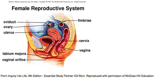
In your Inquiry Into Life textbook on page 420, study the Figure 21.5, The female reproductive system.
The ovaries have two roles in reproduction: to produce egg cells and to produce hormones. Egg production, called oogenesis, takes place in the follicle of the ovary. Eggs are gametes that result from the sexual cell division process called meiosis. This process occurs continuously from puberty to menopause, which occurs at about age 45 to 55.
Eggs are produced cyclically, about once every 28 days. Each time, one or two will eggs mature. At puberty the ovary contains all the primary oocytes (beginning eggs) that a female has. Over time, some oocytes deteriorate, which can lead to genetic abnormalities. The chance of deterioration increases with age.
Hormone production occurs in the developing follicle (mainly estrogen) and the corpus luteum (primarily progesterone). Hormone production is regulated by secretions from the anterior pituitary. This is similar to hormone regulation in males, but is more cyclic, and it is controlled by negative feedback loop.
Eggs are released from the ovary and collected by cilia on the fimbriae. These sweep the eggs into the oviduct (fallopian tube). If missed, the egg could float out into the body cavity. Rarely this leads to ectopic pregnancy where the egg, if fertilized, embeds on the external wall of an organ, usually the fallopian tube. This condition does not lead to proper fetal development.
Once in the fallopian tube, the egg is moved by muscular contractions. This journey lasts for one to two days. It is during this time that the egg must be fertilized for it to develop into an embryo. Eventually the egg enters the uterus, a hollow muscular organ.
The uterus is lined with a spongy tissue, the endometrium. If the egg is fertilized, it embeds itself in the endometrium, which nourishes its early development by providing oxygen and nutrients by diffusion.
The uterus opens into the vaginal canal through an opening called the cervix. Both the uterus and the vagina are very elastic and can stretch to several times their usual size to accommodate a fetus. The vaginal canal opens posterior to the urethral opening and anterior to the anus.
External AnatomyA series of folded tissues, the vulva, collectively surround the vaginal opening. Anterior to the urethral opening is the clitoris. The clitoris contains a collection of nerve endings, similar in number to those in the penis in males. In fact many reproductive structures in males and females have a similar embryonic development, although not necessarily the same function. The following chart summarizes these homologies.

As in males, the development of female secondary sexual characteristics depends on levels of circulating sex hormones. These secondary sexual characteristics include breast development (males have undeveloped breasts), slight deepening of the voice, growth of body hair, muscular development, and a small growth of fatty tissue just below the umbilicus.
Go to the Biology 12 Web site Lesson 43.D Female Reproductive Anatomy and see 2-D and 3-D images/movies of real bodies (male and female) shown as cross-sections. See if you can find parts of the female reproductive system in these pictures.
Go to the Biology 12 Web site Lesson 43.D Female Reproductive Anatomy and see 2-D and 3-D images/movies of real bodies (male and female) shown as cross-sections. See if you can find parts of the female reproductive system in these pictures.
| labelling_the_female_reproductive_system.pdf |
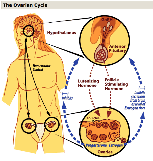
Ovarian and Uterine Cycles
The cycle of female hormone secretions begin at puberty and continue until menopause. With each cycle, the uterus prepares for pregnancy, sloughs off prepared tissue, and prepares again. These cycles are only interrupted by pregnancies or occasional ill health. In this lesson you will learn how some basic hormonal variations influence the ovaries and brain.
The cycle of female hormone secretions begin at puberty and continue until menopause. With each cycle, the uterus prepares for pregnancy, sloughs off prepared tissue, and prepares again. These cycles are only interrupted by pregnancies or occasional ill health. In this lesson you will learn how some basic hormonal variations influence the ovaries and brain.
|
|
Throughout the adult life of the human female, her ovaries respond to neuroendocrine control by undergoing changes that produce eggs and the sex hormones estrogen and progesterone. Feedback to the brain signals the rise and fall of two neuroendocrine hormones, both familiar to you already, LH and FSH. In females these play similar roles as they did in males. FSH stimulates the maturation of the follicle and LH, which stimulates the maturation of the corpus luteum.
Look at Figure 21.7 on page 422 in your Inquiry Into Life textbook, a diagram relating to the ovarian cycle. It shows the events that make up an ovarian cycle, beginning at the top left and ending at the bottom left. In reality, all of these events occur over a period of about one month, and in one place within the ovary. The events numbered 1 to 3 occur in the first fourteen days of the ovarian cycle and those numbered 4 to 6 take place during the last 14 days.
Notice that ovulation—the release of the egg—occurs in the middle of the cycle and is caused by a sudden spike of LH. Some women notice this as a slight pain called Mittelschmerz, from German meaning “middle pain.” Trace the development of the egg inside the primary follicle, to the secondary follicle, and eventually the Graafian follicle. The secondary and Graafian follicles produce the sex hormone estrogen, which is released into the blood stream. After the Graafian follicle ruptures, the empty follicle is known as the corpus luteus (body yellow), named for its colour. The corpus luteum continues to secrete the sex hormone progesterone.
Also in your Inquiry Into Life textbook on page 423, study Figure 21.8, Hormonal control of the ovaries. This diagram is very similar to Figure 21.4 that shows hormonal control of the testes in males. Apart from details of the processes involved in gamete production, they are almost identical. What male hormone is homologous to estrogen? What male hormone is homologous to progesterone?
The Uterine CycleWithin the uterus, conditions change according to secretions of sex hormones from the ovary. While the ovary is involved with egg production, the uterus prepares for the egg’s arrival. The endometrial wall goes through three distinct phases, each corresponding to events within the ovary. Note that the number of days given are averages. The actual length of a cycle varies from woman to woman.
Menstruation
Interrelationships of the Uterine and Ovarian Cycle and Neuroendocrine ControlThe timing of and relationships between hormone levels, ovarian events, and uterine events may seem complex, but it’s easy to understand if we break it down into separate events. Just keep in mind that these events are related to each other, and that they control each other in a negative feedback system.
Take a look at the following diagram of the interrelationships between the ovary, uterus, and brain.
Look at Figure 21.7 on page 422 in your Inquiry Into Life textbook, a diagram relating to the ovarian cycle. It shows the events that make up an ovarian cycle, beginning at the top left and ending at the bottom left. In reality, all of these events occur over a period of about one month, and in one place within the ovary. The events numbered 1 to 3 occur in the first fourteen days of the ovarian cycle and those numbered 4 to 6 take place during the last 14 days.
Notice that ovulation—the release of the egg—occurs in the middle of the cycle and is caused by a sudden spike of LH. Some women notice this as a slight pain called Mittelschmerz, from German meaning “middle pain.” Trace the development of the egg inside the primary follicle, to the secondary follicle, and eventually the Graafian follicle. The secondary and Graafian follicles produce the sex hormone estrogen, which is released into the blood stream. After the Graafian follicle ruptures, the empty follicle is known as the corpus luteus (body yellow), named for its colour. The corpus luteum continues to secrete the sex hormone progesterone.
Also in your Inquiry Into Life textbook on page 423, study Figure 21.8, Hormonal control of the ovaries. This diagram is very similar to Figure 21.4 that shows hormonal control of the testes in males. Apart from details of the processes involved in gamete production, they are almost identical. What male hormone is homologous to estrogen? What male hormone is homologous to progesterone?
The Uterine CycleWithin the uterus, conditions change according to secretions of sex hormones from the ovary. While the ovary is involved with egg production, the uterus prepares for the egg’s arrival. The endometrial wall goes through three distinct phases, each corresponding to events within the ovary. Note that the number of days given are averages. The actual length of a cycle varies from woman to woman.
Menstruation
- days 1 to 5
- wall developed in previous cycle is sloughed off if no fertilized egg is present; should there be a fertilized egg, then the uterine cycle ceases as the embryo develops; this, too, is under hormonal control—read Menstruation on page 424 of the Inquiry Into Life textbook
- often associated with pain—read Painful Menstruation and Premenstrual Syndrome on page 424 of the Inquiry Into Life textbook
- days 6 to 13
- endometrial wall thickens as it develops new tissue and blood vessels
- ends with ovulation, day 14-for an embryo to develop, the egg must be fertilized within a couple of days of ovulation
- days 15 to 28
- secretes mucous, which accumulates in strands inside the uterus; these provide a pathway for sperm that must swim through the uterus and into the fallopian tubes to meet with the egg. Later the mucous traps the fertilized egg, which eventually embeds into the uterine wall. If the egg is not fertilized, it degenerates.
- without a fertilized egg, the endometrium begins to degenerate and the cycle begins at day one again.
Interrelationships of the Uterine and Ovarian Cycle and Neuroendocrine ControlThe timing of and relationships between hormone levels, ovarian events, and uterine events may seem complex, but it’s easy to understand if we break it down into separate events. Just keep in mind that these events are related to each other, and that they control each other in a negative feedback system.
Take a look at the following diagram of the interrelationships between the ovary, uterus, and brain.
|
|
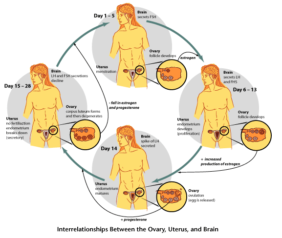
|
Follow the arrows through each cascade of events. Notice that events in one organ control those in the others. Hormones are the communicators. Then take a look at Figure 21.9 on page 425 in the Inquiry Into Life. The diagram in the Inquiry Into Life textbook does not describe the controls, so be sure to study the one provided here in the lesson. On the provincial exam, students are often asked questions about the control mechanisms.
Watch The Female Cycle now.
Female Cycle Animation
Watch The Female Cycle now.
Female Cycle Animation